Page Contents
Features of Photographic Atlas for Anatomy & Physiology PDF
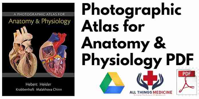
A Photographic Atlas for Anatomy & Physiology PDF is a new visual lab study tool that helps students learn and identify key anatomical structures.
Featuring photos from Practice Anatomy Lab™ 3.0 and other sources, the Atlas includes over 250 cadaver dissection photos, histology photomicrographs, and cat dissection photos plus over 50 photos of anatomical models from leading manufacturers such as 3B Scientific®, SOMSO®, and Denoyer-Geppert Science Company.
The Atlas is composed of 13 chapters, organized by body system, and includes a final chapter with cat dissection photos. In each chapter, students will first explore gross anatomy, as seen on cadavers and anatomical models, and then conclude with relevant histological images.
Recommended Books For You
 General Surgery Coding 2020 Edition PDF
 AMC Handbook of Clinical Assessment Pdf
Description of Photographic Atlas for Anatomy & Physiology PDF
Photographic Atlas for Anatomy & Physiology PDF is one of the best-known books on the subject of basic medical sciences. This book covers all the cases and phenomenons a student and professional doctor might be up against in their whole life. Master this book and you will be of prime help in solving cases of diseases that are difficult to treat. Make a difference. Download Now.
The Authors
Nora Hebert, Ph.D. teaches undergraduate courses in Anatomy and Physiology at Red Rocks Community College near Denver, Colorado. Although most of her students are undergraduates, primarily interested in the allied health professions, Nora has also taught graduate-level Human Physiology for the College’s Physician Assistant Program.
Nora is an active faculty member at Red Rocks, serving on the faculty senate, the honors program committee, and the admissions and executive committees for the Physician Assistant Program. She is also part of the College’s Campus Green Initiative.
Among her many academic projects, Nora has consulted in the development of an interactive virtual knee, known as the Explorable Virtual Human, with the Center for Human Simulation at the University of Colorado Health Sciences Center. She has also been involved with the Visible Human Dissector program, advising K-12 teachers and postsecondary instructors on how best to implement the Dissector in their classrooms. Nora has been deeply involved in the development of Practice Anatomy Lab, as coauthor of versions 2.0 and 3.0. She is also the author of over 60 A&P Flix animations covering muscle physiology, neurophysiology, and muscle origins, actions, insertions, and innervations.
Nora received a Ph.D. in Endocrinology from the University of California at Berkeley.
Ruth E. Heisler is a senior instructor in the Department of Integrative Physiology at the University of Colorado at Boulder where she teaches and coordinates several courses, including Human Anatomy, Comparative Vertebrate Anatomy, and Forensic Biology. She has been an instructor at the University of Colorado for more than 15 years.
At the University of Colorado, Ruth has worked extensively with the Science Education Initiative to improve both the teaching and understanding of scientific material at the undergraduate level. In addition, she has been involved in academic outreach through workshops with the American Academy of Forensic Sciences and the Biological Sciences Initiative. She has been a consultant on projects with the Center for Human Simulation, working with data generated through the Visible Human Project.
Ruth has been deeply involved in the development of Practice Anatomy Lab, as coauthor of versions 2.0 and 3.0. She is also author of a custom laboratory manual developed for a large, cadaver-based human anatomy lab.
Ruth received her B.S. in Biology from the University of Minnesota, and her M.A. in Biology from the University of Colorado.
Jett Chinn is an instructor of Human Anatomy in the Science and Technology Division of Cañada College (Redwood City, CA) and also the Life and Earth Sciences Department at the College of Marin (Kentfield, CA).
Jett has more than 20 years of experience teaching Human Anatomy at institutions including San Francisco State University, California College of Podiatric Medicine, and Touro University College of Osteopathic Medicine. He has also taught first-year dental students at the UC San Francisco School of Medicine.
Jett received a B.A. in general biology from San Francisco State University.
Karen M. Krabbenhoft, Ph.D. is a senior lecturer in the Department of Neuroscience at the University of Wisconsin in Madison. During her 20-year career, Karen’s focus has been on teach
Dimensions and Characteristics of Photographic Atlas for Anatomy & Physiology PDF
- Publisher : Pearson; 1st edition (October 14, 2014)
- Language : English
- Loose Leaf : 240 pages
- International Standard Book Number-10 : 0321869257
- International Standard Book Number-13 : 978-0321869258
- Item Weight : 3.53 ounces
- Dimensions : 8.6 x 0.7 x 10.95 inches
- Book Name Photographic Atlas for Anatomy & Physiology PDF
Download Link 1
Top reviews
I have to retake one of my A&P classes to get a ‘nursing school application-appropriate-grade’, and honestly if I had purchased this sooner, I probably wouldn’t have to be in a position to retake it because everything you need to master in lab is in this book!
If you are starting A&P to get into a health program, you need to have this with you. The anatomical models in this book are the EXACT models we are tested on. The human cadaver photos are really well done, with great specimens to reference to. I’m so frustrated I didn’t have this when I FIRST started my journey, but at least I have this now!“
The atlas has three holes — meant to go into a 3-ring binder. I didn’t intend to order this type — I wanted a manual without holes (and thought I ordered this). But it worked out better this way because now it’s easier to take out individual pages from the binder to show to my students. LOVE this atlas!”

Disclaimer:
This site complies with DMCA Digital Copyright Laws. Please bear in mind that we do not own copyrights to this book/software. We’re sharing this with our audience ONLY for educational purposes and we highly encourage our visitors to purchase the original licensed software/Books. If someone with copyrights wants us to remove this software/Book, please contact us. immediately.
You may send an email to emperor_hammad@yahoo.com for all DMCA / Removal Requests.

